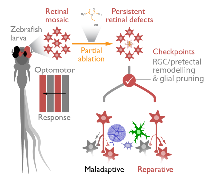
Background: Retinal cell loss due to injury or disease results in compromised visual input to the brain and triggers circuit remodelling in central visual circuits.

Hypothesis: Remodelling in central visual circuits involving retinal ganglion cell axons and pretectal neurons represents an important compensatory checkpoint for the recovery of visual function.

Strategy: We will ablate red cones in the retina, probe changes in network activity and structural remodelling in central visual circuits and investigate the contribution of glial cells to this synaptic plasticity to reveal the mechanisms mediating the recovery of visual function.

Summary

The circuit remodelling that occurs in many paradigms of brain trauma and disease, is thought to represent compensatory network changes that play an important role in the restoration of lost function. We have been investigating network remodelling in the visual system of larval zebrafish following acute injury in the retina, as this vertebrate model organismlends itself to an unbiased, systems-based approach to identify the mechanisms of circuit repairthat ultimately lead to functional recovery.Our approach is simple: we ablate red cone photoreceptors in the retina using chemogenetic tools and use behavioural assays as a functional readout of the brain network. As a consequence of the induced retinal cell loss, we observed initial behavioural visual deficits that subsequently recovered. This restoration of visual function occurred despite the incomplete replacement of ablated retinal cells. Moreover, whole-brain calcium imaging experiments revealed that behavioural recovery occurred contemporaneously with adaptive network changes in the pretectum, an early visual center. The pretectum, which is analogous to the nucleus of the optic tract in mammals, is one synapse downstream of the retinal ganglion cell (RGC) axons that convey visual input from the retina to the central brain, and it is known to be involved in processing the type of visual stimulus we presented. These experiments lead to the clear hypothesis that the RGC to pretectum connectivity undergoes structural and/or functional rewiring during network compensation. In the second funding period we will therefore investigate the structural and/or functional changes in central circuits, involving RGC axons and pretectal neuron dendrites in Aim ❶. In particular, we will investigate three (non-exclusive) options (i) RGC axons compensate by altering their innervation onto pretectal dendrites, (ii) pretectal neurons compensate for the decreased visual input by altering their dendritic trees and (iii) neuronal morphology, at the level of the processes, remains the same but connectivity becomes stronger through an increase in the number of synapses or their strength. Given the well-recognized roles of microglia, astroglia and more recently oligodendrocyte precursor cells in synaptic remodelling, we will investigate the role of glial cells in remodelling RGC axon-pretectal connectivity in Aim ❷. We hypothesize that at least one of the glial cell-types will play a central role in the structural remodelling of these central circuits.  Once we identify the primary glial effector of remodelling, we will assess the underlying molecular mechanisms involved in this process.