Background: Childhood-onset white matter diseases (WMDs) are characterized by inflammatory tissue damage.

Hypothesis: CD8+ T cell pheno-types within demyelinating brain lesions are tipping the balance towards damage or repair.

Strategy: Identification and visualization of CD8+ T cell-driven disease and/or repair pathways with multimodal approaches combining multifluorescent immunohistochemistry, spatial transcriptomics, serum biomarkers and MR imaging.

Summary

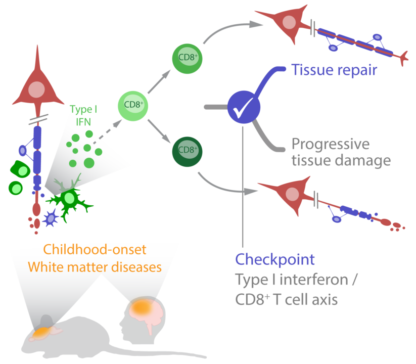
Childhood-onset white matter diseases including multiple sclerosis (MS) and monogenetic leukoencephalopathies such as X-linked adrenoleukodystrophy (X-ALD) and RNAseT2 deficiency share CD8+ T cell-dominated neuroinflammation, demyelination, axonal damage, and variable oligodendrocyte pathology. Disease course, therapy response and spontaneous recovery vary greatly within and between these diseases. A differentiated understanding of the drivers leading to damage and repair is an unmet need of high clinical relevance. 

The type I interferon (IFN)– CD8+ T cell axis is an important immune checkpoint in inflammatory leukoencephalopathies, as we recently demonstrated in a new mouse model. Spatial transcriptome analysis of MS lesions suggests that this checkpoint is also relevant to other inflammatory white matter diseases. Therefore, we propose that CD8+ T cell differentiation dynamics, influenced by type I IFN, have a profound impact on disease manifestation and progression in childhood onset monogenetic and autoimmune inflammatory white matter diseases.In the second funding period, we will provide a lesion stage-dependent characterization ofCD8+ T cell subpopulations across the white matter disease spectrum. Effector mechanisms of CD8+ T cells will be defined by integrated single cell and spatial transcriptome analysis, and the role of specific CD8+ T cell subpopulations will be examined. Furthermore, we will dissect the contribution of type I IFN and CD8+ T cells to lesion development, progression, and recovery in cell type-specific knockout mice and by antibody-mediated cell depletion strategies. The lesion patterns and mechanisms identified in the animal model will be correlated with the patient phenotypes through human tissue analysis, serum biomarker studies and MRI analysis. The knowledge gained about the checkpoints will be used to establish early disease markers and to identify new therapeutic targets that influence the survival of axons and oligodendrocytes and improve myelin repair.