Background
In Multiple Sclerosis (MS), focal lesions and white matter pathology have been long studied. Grey matter changes however, particularly in the cortex, are increasingly recognised as strong predictors of progression, disability and cognitive decline. In-vivo detection of cortical synapse loss, an early indicator of grey matter pathology, has remained challenging.
Aims
The study set out to test whether positron emission tomography (PET) imaging of the synaptic vesicle protein 2A (SV2A), using the radiotracer [¹⁸F]UCB-H, can detect synaptic loss in cortical grey matter in MS, first in a mouse model, then in human patients, and whether the imaging signal correlates with synapse density and clinical measures of disease.
Key Findings
- The authors confirmed that SV2A is a valid marker for synaptic density in MS cortical grey matter via tissue analysis of SV2A mRNA and protein.
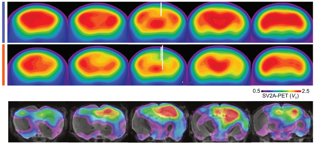
- In a mouse model of cortical MS pathology, SV2A-PET imaging detected synapse loss in cortical lesions, and the PET tracer uptake estimates corresponded with immunohistochemically measured synapse densities in the same lesions.
- In a cohort of 31 individuals with MS at various disease stages, SV2A-PET revealed cortical synaptic abnormalities in vivo. Notably, hemispheric asymmetries in tracer uptake pointed to cortical pathology beyond what traditional MRI white-matter imaging detects. The volume of PET-defined cortical synapse loss was more than 20-fold larger than cortical lesion volume detected with MRI in the same participants.
- PET-defined cortical synapse pathology showed relationships with greater disability, cognitive deficit and fatigue. In particular, more extensive synaptic loss correlated with more advanced / progressive disease stages.
Implications
- SV2A-PET provides a new way to visualise and quantify cortical synapse loss in MS, a lesion pathology which has been elusive in vivo until now.
- Because synaptic loss likely underlies irreversible neural network damage and cognitive decline in MS, having a tool to detect it opens up possibilities for earlier diagnosis of disease progression and monitoring of neuroprotective treatments.
- The fact that PET-derived synapse-loss volumes far exceed MRI-detected cortical lesion volumes suggests that current imaging may underestimate cortical pathological burden; this could influence how clinical progression is interpreted and how treatments are stratified.
- Long-term, this method might help in clinical trials to assess the efficacy of therapies aimed at preventing synapse loss or restoring synaptic integrity in MS.
Conclusion
This translational study successfully demonstrates that SV2A-PET imaging can detect and quantify cortical synapse loss in multiple sclerosis both in preclinical models and in patients. The strong correlation with histology and clinical measures suggests that cortical synaptic pathology is more widespread than visible with MRI and is meaningful for functional outcomes. The work stands to advance the monitoring of grey matter pathology in MS and may help shift focus toward synapse-preserving therapeutic strategies.
📖 Full text for further details: Science Translational Medicine
TRR 274 Project leaders contributing to this project: Martin Kerschensteiner (B07 & C02).